A groundbreaking study from Zhongshan Hospital has unlocked the long-standing “black box” of how liver cancer evades immune surveillance and spreads to the lungs. Published in Science, the research reveals the complete spatiotemporal immune escape mechanism behind hepatocellular carcinoma (HCC) lung metastasis, offering precise timing and targets to block tumor spread.
Lung metastasis is the most common distant spread of liver cancer, affecting up to 55% of patients with extrahepatic metastasis. Most disseminated tumor cells (DTCs) are rapidly eliminated by the lung’s immune system after entering circulation. Only a tiny fraction survives dormancy, grows into microscopic lesions, and eventually develops into fatal macroscopic metastasis. Due to its invisibility in clinical imaging, the full evolutionary process of early tumor immune escape has remained unclear for decades.
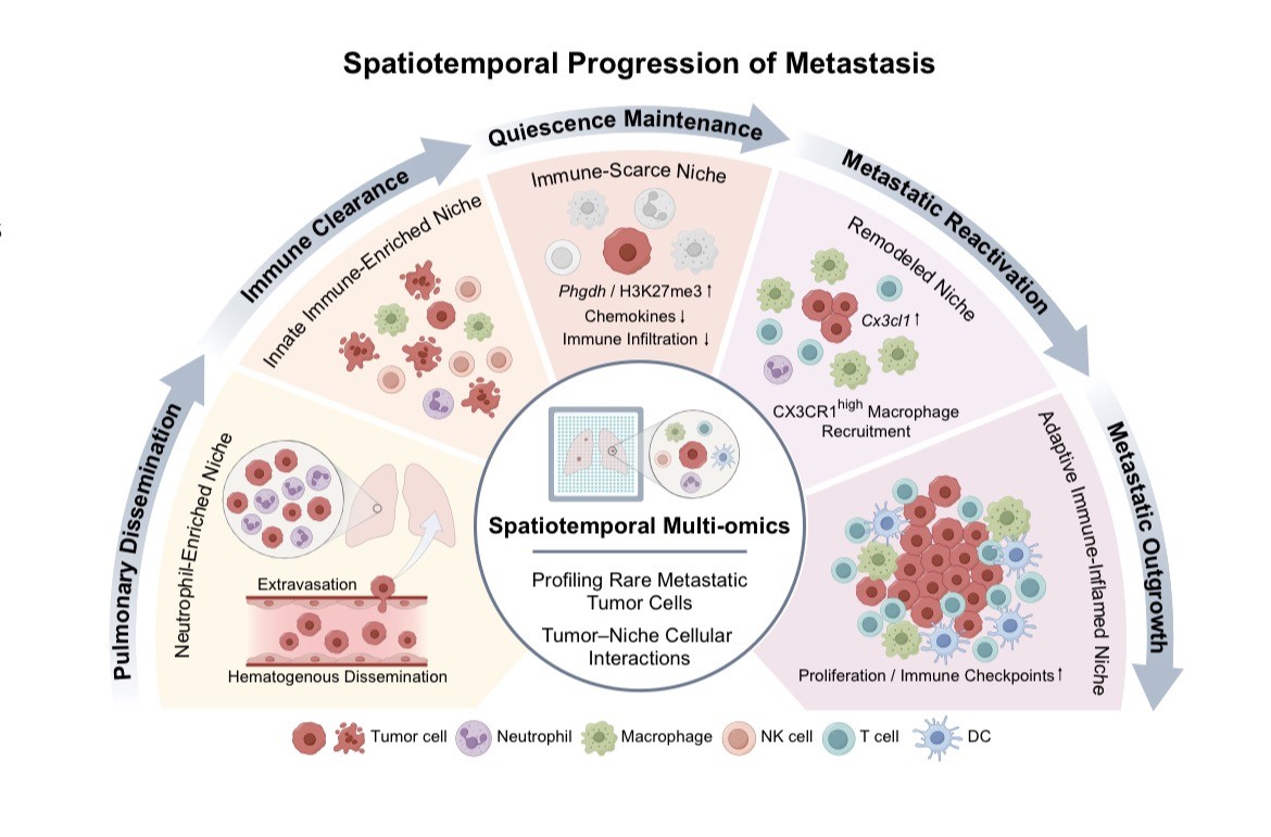
To solve this puzzle, the research team adopted high-precision spatiotemporal multi-omics technology and constructed a full-stage dynamic atlas covering nine consecutive time points. For the first time, the study fully reproduces the complete metastatic journey: tumor cell dissemination, immune dormancy, reactivation, and macroscopic colonization. It further divides lung metastatic colonization into four sequential stages and clarifies the bidirectional evolutionary relationship between tumor cells and the lung microenvironment.
Different from traditional single-gene linear research, this study confirms that cancer metastasis is not driven by one-way tumor genetic changes. Instead, it is shaped by continuous crosstalk between tumor cells and host tissues. Alveolar epithelial cells induce tumor cell dormancy, while surviving cancer cells reshape the lung into an immunosuppressive “immune desert”. In the transition stage from microlesion to overt metastasis, tumors recruit CX3CR1⁺ macrophages, which further gather immune-suppressive cells and create favorable conditions for tumor growth.
The research also upgrades the traditional static tumor analysis model. By tracking continuous temporal and spatial changes, it transforms metastasis research from “snapshot observation” to “dynamic ecological simulation”. Clinically, the study delivers a key insight: treatment targets and intervention timing are equally critical.
Researchers identified that high PHGDH expression is a transient adaptive state of surviving tumor cells, rather than a fixed genetic feature. Most fatal metastatic cells experience high PHGDH activity at the early dormant stage, which declines once tumors expand. Targeted intervention during this critical adaptive window — including epigenetic regulation to reverse the immune desert or macrophage clearance during micro-metastasis transition — can effectively block metastasis, while treatment at other stages yields limited effect.
This landmark study redefines cancer metastasis as a spatiotemporal ecological dynamic process, rather than a simple linear genetic cascade. It establishes PHGDH and CX3CR1⁺ macrophages as novel precise intervention targets and proposes a new “target plus timing” dual-dimensional strategy for anti-metastasis therapy.
The findings fill the clinical gap in early occult micro-metastasis intervention, provide a comprehensive theoretical basis for staged anti-recurrence management, and open a new precision pathway to block distant spread for liver cancer and other malignant tumors worldwide.